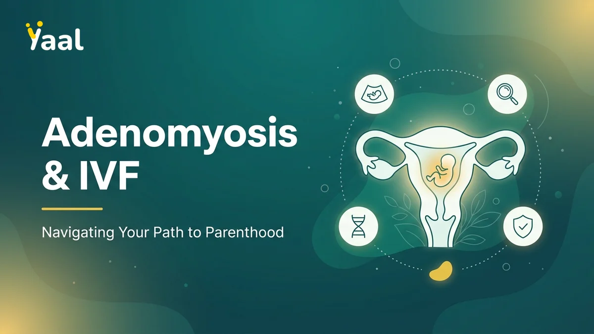
For many women struggling to conceive, understanding the complex relationship between adenomyosis and fertility is the first step toward finding a clear, evidence-based path to parenthood. While this uterine condition presents distinct anatomical and physiological challenges, advanced reproductive technologies now offer highly successful pathways to pregnancy. By exploring how adenomyosis impacts the uterus and learning about modern therapeutic strategies, you can make informed decisions alongside your fertility specialist.
What is Adenomyosis and How Does It Differ From Endometriosis?
Adenomyosis is a progressive gynecological condition characterized by the abnormal presence of endometrial tissue—which normally lines the inside of the uterine cavity—deep within the muscular wall of the uterus, known as the myometrium. Each month, during the menstrual cycle, this misplaced tissue responds to hormonal signals by thickening, breaking down, and bleeding. Because this blood has no way to escape the muscular wall, it pools inside the myometrium, causing localized inflammation, swelling, and the formation of fibrous tissue. Over time, this process leads to an enlarged, globular, and hardened uterus that cannot function optimally.
Adenomyosis vs. Endometriosis: Key Differences
While adenomyosis and endometriosis are sister conditions sharing similar cellular characteristics, they are anatomically distinct.
- Adenomyosis: The endometrial-like tissue is confined entirely within the muscular wall of the uterus (myometrium). The uterus itself becomes bulky, heavy, and structurally compromised.
- Endometriosis: The endometrial-like tissue grows outside the uterine cavity entirely. It is commonly found on the ovaries (forming chocolate cysts or endometriomas), fallopian tubes, outer uterine wall, pelvic ligaments, or even the bowels and bladder.
This anatomical distinction is crucial for fertility planning, as the two conditions alter reproductive physiology in entirely different ways. Endometriosis primarily impacts egg quality and pelvic anatomy through adhesions, whereas adenomyosis directly compromises the structural integrity and receptivity of the womb itself.
Can You Have Both Conditions Simultaneously?
Yes, adenomyosis and endometriosis frequently coexist. Clinical studies indicate that a significant percentage of women diagnosed with severe endometriosis also have varying degrees of adenomyosis. When both conditions are present, they can create a compounding effect on pelvic pain and reproductive challenges. The overlapping symptoms—such as severe menstrual cramps and deep pelvic pain—often delay a precise diagnosis. Managing coexisting cases requires a highly integrated, multi-phased treatment protocol that addresses both the pelvic inflammation caused by endometriosis and the structural uterine changes caused by adenomyosis.
How Adenomyosis Affects Fertility and Embryo Implantation
The presence of ectopic endometrial tissue within the myometrium alters the biological, chemical, and physical environment of the uterus. For a successful pregnancy to occur, several highly coordinated steps must take place: sperm must migrate through the uterus, the fallopian tubes must capture the egg, and the fertilized embryo must find a receptive endometrial lining to implant. Adenomyosis disrupts this delicate sequence at multiple points.
Impact on the Endometrial Lining
The chronic, localized inflammation within the myometrium spills over into the endometrium (the inner lining). This inflammatory microenvironment elevates levels of free radicals and inflammatory cytokines, which are toxic to developing embryos. Furthermore, adenomyosis is associated with a reduction in key implantation markers, such as HOXA10 genes, and a state of progesterone resistance. Progesterone is the primary hormone responsible for preparing the endometrium for implantation. When the lining becomes resistant to progesterone, it fails to undergo the necessary structural changes (decidualization) required to welcome and nourish an incoming embryo.
Uterine Contractility and Sperm Transport
A healthy uterus exhibits subtle, rhythmic waves of muscle contractions that change direction depending on the phase of the menstrual cycle. During ovulation, these contractions gently direct sperm upward toward the fallopian tubes. In a uterus affected by adenomyosis, the normal architecture of the junctional zone—the specialized boundary layer between the endometrium and myometrium—is disrupted. This disruption leads to dysregulated, hyperactive, or uncoordinated uterine contractions. These abnormal spasms can actively hinder sperm transport, preventing fertilization from occurring naturally. Additionally, these hyper-contractions can physically disrupt a newly arrived embryo, preventing it from adhering to the uterine wall.
Recognizing the Symptoms of Adenomyosis

The clinical presentation of adenomyosis varies widely from woman to woman. While some experience debilitating physical symptoms, others remain entirely unaware of the condition until they face difficulties conceiving. Recognizing these indicators early can significantly reduce the time spent struggling with unexplained infertility.
Common Signs to Watch For
The most characteristic symptoms of adenomyosis stem from the swelling and bleeding occurring within the uterine muscle:
- Menorrhagia: Exceptionally heavy or prolonged menstrual bleeding, often accompanied by the passage of large blood clots.
- Dysmenorrhea: Severe, throbbing, or cramping pelvic pain during menstruation that often worsens over time and is poorly responsive to standard over-the-counter pain relievers.
- Dyspareunia: Pain during or after sexual intercourse, caused by the inflammation and tenderness of the enlarged uterine body.
- Chronic Pelvic Pain: A persistent, dull ache in the lower abdomen or lower back that remains present throughout the menstrual cycle.
- Abdominal Bloating: A sensation of pressure or fullness in the lower abdomen, often described as feeling like the abdomen is hard or visibly distended.
The Reality of Silent Adenomyosis
It is a common misconception that adenomyosis always presents with heavy bleeding and pain. A notable subset of patients experiences "silent" adenomyosis. In these cases, the disease may be focal (restricted to a small area of the uterine wall) or slowly progressing, causing no noticeable daily symptoms.
For these women, the primary symptom is the inability to conceive or experiencing recurrent early pregnancy losses. This highlights the vital importance of comprehensive reproductive imaging during the initial stages of any fertility assessment, even if a woman has regular, painless menstrual cycles.
How is Adenomyosis Diagnosed for Fertility Planning?
Historically, adenomyosis could only be definitively diagnosed after a hysterectomy by examining the uterine tissue under a microscope. Today, advanced non-invasive imaging technologies allow fertility specialists to accurately diagnose and map the extent of the disease, which is essential for tailoring an effective conception strategy.
Advanced Ultrasound Techniques
High-resolution Transvaginal Ultrasound (TVS) serves as the primary, highly accessible screening tool for adenomyosis. When performed by an experienced reproductive sonographer, TVS can identify several key markers of the condition, including:
- An asymmetrical thickening of the anterior or posterior uterine walls.
- The presence of small, fluid-filled myometrial cysts.
- A "shaggy" or poorly defined endometrial-myometrial junction.
- An overall globular, enlarged appearance of the uterus without the presence of distinct fibroids.
- Increased vascularity (blood flow) within the affected areas of the myometrium.
When is a Pelvic MRI Recommended?
While ultrasound is an excellent initial tool, a Pelvic Magnetic Resonance Imaging (MRI) is considered the gold standard for detailed fertility planning. An MRI provides highly detailed, multi-planar images of the pelvic anatomy. It is particularly recommended when:
- Ultrasound findings are inconclusive or suspect co-existing uterine fibroids.
- The specialist needs to precisely measure the thickness of the junctional zone (a junctional zone thickness of 12mm or greater is highly indicative of adenomyosis).
- The clinical team is planning a uterus-sparing surgical intervention and requires an exact map of the adenomyotic lesions versus healthy muscle tissue.
Hysteroscopy may also be utilized alongside these imaging modalities. While a hysteroscopy cannot see inside the uterine wall itself, it allows the specialist to view the internal cavity directly, ruling out concurrent issues like endometrial polyps or intrauterine adhesions before beginning fertility protocols.
Medical and Surgical Treatment Options for Adenomyosis
Managing adenomyosis in patients who still wish to conceive requires a delicate, highly specialized approach. Standard treatments for non-fertility patients, such as a hysterectomy or continuous oral contraceptives, are not suitable for those actively trying to achieve pregnancy. Instead, therapies must focus on temporarily suppressing the disease or conservatively removing it to preserve uterine function.
Hormonal Suppression Therapy
Because adenomyosis is an estrogen-dependent condition, temporarily depriving the abnormal tissue of estrogen can cause it to shrink and quiet down.
- GnRH Agonists: Medications such as leuprolide acetate are commonly used to induce a temporary, reversible state of menopause. By shutting down ovarian estrogen production for 2 to 3 months, these medications allow adenomyotic lesions to shrink, drastically reducing localized uterine inflammation and restoring the endometrial lining to a more receptive state.
- Anti-inflammatory Regimens: High-dose, targeted non-steroidal anti-inflammatory drugs (NSAIDs) are often prescribed during active cycles to lower prostaglandin levels in the uterus, mitigating both pain and abnormal uterine contractions.
Uterus-Sparing Surgical Interventions
In cases of focal adenomyosis (where the disease is concentrated in one defined area, often called an adenomyoma), conservative surgery may be considered.
- Adenomyomectomy: This specialized surgical procedure aims to excise the diseased, adenomyotic tissue while carefully reconstructing the healthy muscular wall of the uterus. Because adenomyosis does not have clear margins like fibroids do, this surgery requires exceptional surgical skill to ensure the uterine wall remains strong enough to safely carry a future pregnancy.
- Post-Surgery Recovery: Following an adenomyomectomy, a healing period of several months is mandatory before attempting pregnancy or embryo transfer to ensure complete structural healing of the myometrium.
Optimizing IVF Success Considerations with Adenomyosis
In vitro fertilization (IVF) is often the most effective route to pregnancy for women with moderate to severe adenomyosis. However, standard IVF protocols must be carefully modified to address the specific challenges posed by an inflamed uterine environment.
Why Frozen Embryo Transfer (FET) is Key
In a standard "fresh" IVF cycle, high doses of stimulating hormones can cause estrogen levels to skyrocket. In a patient with adenomyosis, this sudden surge of estrogen can cause the adenomyotic lesions to flare up, leading to acute uterine inflammation and hyper-contractility at the exact moment of embryo transfer.
To avoid this, a Frozen Embryo Transfer (FET) strategy is highly preferred. In an FET cycle:
- Oocytes are retrieved and fertilized in the laboratory.
- The resulting high-quality embryos are safely cryopreserved (frozen).
- The patient's uterus is allowed to rest and return to a natural, calm state.
- The transfer is scheduled for a subsequent, controlled cycle where the uterine lining can be prepared optimally without the hyper-stimulation of the ovaries.
Pre-Transfer Downregulation Protocols
To maximize the success of an FET, specialists often employ a "prolonged downregulation" protocol. This involves administering a GnRH agonist for 2 to 3 months immediately prior to the scheduled embryo transfer.
| Phase | Duration | Clinical Objective |
|---|---|---|
| 1. Downregulation | 8 to 12 Weeks | Suppress estrogen, shrink adenomyotic tissue, and eliminate uterine inflammation. |
| 2. Endometrial Preparation | 2 to 3 Weeks | Administer controlled estrogen and progesterone to build a receptive, uniform lining. |
| 3. Embryo Transfer | Single Day | Place a high-quality thawed embryo into a calm, receptive, and non-inflamed uterus. |
This customized approach dramatically improves implantation rates by ensuring that the embryo is introduced into an environment that mimics a healthy, non-adenomyotic womb as closely as possible.
Expert Adenomyosis and Fertility Care in Chennai
Overcoming the challenges of adenomyosis requires a combination of cutting-edge reproductive technology, clinical expertise, and deeply compassionate care. At Yaal Fertility Centre, we specialize in helping couples navigate complex uterine and reproductive conditions with highly individualized treatment pathways.
Personalized Care at Yaal Fertility Centre
Every patient's journey with adenomyosis is unique. At our state-of-the-art facility in Chennai, we do not believe in one-size-fits-all protocols. We combine high-resolution pelvic imaging, detailed hormonal profiling, and advanced embryology services to design a treatment roadmap tailored specifically to the severity of your condition, your age, and your family-building goals. Our clinical philosophy focuses on patient comfort, clear communication, and clinical excellence.
Consulting Dr. Rukkayal Fathima
Led by the esteemed Dr. Rukkayal Fathima, MRCOG (UK), our medical team brings world-class fertility training and international standards of care to Chennai. Dr. Fathima's extensive experience in managing complex uterine factors, endometriosis, and recurrent implantation failures ensures that patients with adenomyosis receive the most advanced, evidence-based care available.
By utilizing advanced pre-transfer downregulation protocols, meticulous frozen embryo transfer techniques, and customized hormonal support, our clinical team consistently achieves an outstanding 70% IVF success rate, bringing hope and healing to families.
Frequently Asked Questions About Adenomyosis and Fertility
Can I get pregnant naturally if I have adenomyosis?
Yes, natural pregnancy is entirely possible, especially in mild or focal cases of adenomyosis. However, if you have been actively trying to conceive for over six months (if over age 35) or a year (if under age 35) without success, it is advisable to consult a fertility specialist to assess if the condition is impacting your uterine receptivity.
Does adenomyosis increase the risk of miscarriage?
Yes, studies indicate that the localized inflammation and altered blood flow associated with adenomyosis can modestly increase the risk of early pregnancy loss. Fortunately, preparing the uterine lining with temporary hormonal downregulation prior to conception or embryo transfer significantly mitigates this risk.
How does a doctor tell the difference between adenomyosis and fibroids?
While both conditions cause an enlarged uterus and heavy bleeding, fibroids are benign tumors of well-defined muscle tissue that can be easily "peeled" away from the healthy uterus. Adenomyosis, conversely, consists of diffuse, poorly defined endometrial tissue scattered directly throughout the normal uterine muscle fibers, which is clearly visible on high-resolution MRI scans.
Will treating my adenomyosis cure my pelvic pain?
Yes, therapies designed to suppress adenomyosis for fertility—such as GnRH agonists—typically provide substantial relief from pelvic pain, heavy bleeding, and cramping during the treatment period by temporarily shrinking the inflamed tissue.
Taking Your Next Steps Toward Parenthood
While a diagnosis of adenomyosis can feel overwhelming, it is important to remember that it is a manageable condition. With the advent of advanced diagnostic imaging, prolonged hormonal downregulation, and highly refined frozen embryo transfer protocols, the barrier that adenomyosis once posed to pregnancy has been significantly lowered. Early, proactive evaluation is the key to preserving your reproductive options and avoiding unnecessary delays on your path to parenthood.
If you are experiencing symptoms of adenomyosis or have faced difficulties conceiving, taking the next step with an experienced specialist can make all the difference. Book an appointment with Yaal Fertility Centre today. Rukkayal Fathima and our dedicated fertility team in Chennai.
Frequently Asked Questions
Written by
Dr. Rukkayal Fathima
Specialist at Yaal Fertility Centre, Chennai. Providing expert guidance on fertility and women's health.